Tibial Hemimelia (TH) in Cattle
This was an old report I did for my Farm Animal Anatomy and Physiology class when I was in college and this got a pretty dang good grade in that class (I was struggling in that class). So now, I present it here because it has excellent and educational information about Tibial Hemimelia (TH) in cattle. Along with that, it's something to consider if you're interested in breeding cattle.
Introduction
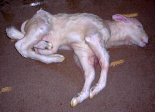 |
| A calf born with Tibial Hemimelia (photo from steerplanet.com) |
History
Tibial hemimelia was first described in the 1950s, in Scotland (Young, 1951). It has also been described in Canada (Lapointe, Lachance, and Steffen, 2000) and the United States (Whitlock, Kaiser, and Maxwell, 2008). While it is definitely possible that it has been seen many times, it was most likely unreported due to that the owners of the calves’ mothers believed that it was a physical defect and nothing else. This idea was overturn when TH was first described in 1951 and it has been suggested that TH was caused by a mutation (Young, 1951). Several decades later, it has been proven that TH is genetic and that the original mutation in the Shorthorn breed has been traced to a bull from Ireland, that goes by the name of Deerpark Improver (Whitlock, Kaiser, and Maxwell, 2008), which was a popular breeding sire used during the 1970s. A result of using this shorthorn bull was the occurrence of TH has been increasing quite dramatically.
Description
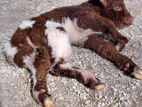 |
| Red-and-White Shorthorn calf with TH (photo from steerplanet.com) |
As I mentioned earlier, TH is caused by a genetic defect (and not by any other environmental factors), a mutation within the sire which is seen in several individuals of many breeds (Whitlock, Kaiser, and Maxwell, 2008). It is said to be more common in certain breeds, such as Shorthorn (Marron, Thurnau, Hannon, and Beever, 1974) and Galloway cattle (Ojo et al. 1974), due to continuous inbreeding and line-breeding (Lapointe, Lachance, and Steffen, 2000). While this mutation has caused similar symptoms in the calves, the amount of similar symptoms do vary between breeds. What has caused this mutation is an autosomal recessive allele in the genetic material that is responsible for the regulation of the formation of the hind legs (Whitlock, Kaiser, and Maxwell, 2008).
To understand about how the animal is either a carrier or a non-carrier and to understand how an animal could carry the gene for TH and not show it is by understanding simple principles of inheritance. If an animal is carrying the gene for TH, but doesn’t show that it has TH, that animal would be heterozygous. What this means is that in the animal’s genetic material, it carries the recessive trait for TH (t), but as the dominant trait that cancels out the recessive trait’s expression (T). To put it simply, we would say that the animal’s alleles were something like this symbol (Tt), it will carry the recessive trait for TH but it will also carry the dominant trait that inhibits the recessive one. Along with that, we would say the animal that does not carry the recessive trait is homozygous, which would mean that all its alleles have only dominant traits (TT). At the same time, the calf that is expressing the recessive TH trait would be called homozygous as well (tt). But, since that calves that do express the recessive trait never live up to sexual maturity or even survive birth, the only way to pass down the trait is through the heterozygous animals or the carriers. If the heterozygous carriers have mated with the homozygous non-carriers, then about half of their halves will become carriers of the TH trait (Tt) and another half that will be homozygous dominant. But if a heterozygous carrier has mated with another heterozygous carrier, then you would have calves that would express the TH trait (tt), most will be carriers (Tt), and some normal calves (TT) born. How the mutation affects the calf is that during the very early stages of development, the TH mutation removes a relatively large part of the gene that is responsible for the formation of the hind legs (Whitlock, Kaiser, and Maxwell, 2008), which in turn, causes a large multitude of negative traits to arise.
Since I have described the genetic component of the condition, let me explain the physical aspect of the condition. As mentioned earlier, the tibia in the hind limbs of the TH calf would be in a corkscrew shape. But this is not the only characterized trait in TH, there several more traits to come. Another skeletal trait of TH is the abnormal structure of the distal femoral epiphysis (Guffy and Leipold, 1977), which is the part of the femur bone that meets with the tibia. One other set of traits would be a retained set of testicles (which is called cryptorchidism) or the absence of one in the case of male calves and failed mullerian duct (which is reasonable for the development of the female’s reproductive organs, such as the uterus and fallopian tubes) development is the case of female calves. Another trait that comes with TH is meningocele, which is a genetic defect which causes the embryonic neural tube of the calf to be either not close or close up too much or too soon. Along with that trait, another one would be a large abdominal hernia (when a hole develops in a membrane, it allows an organ or organs to stick through). One other trait the does appear in TH calves is that the skull of the resulting calf is not enclosed and that the brain would be exposed. While I am on the subject of the skull, there is a trait or two that affects the brain as well. One such trait would be that the brain would be missing the septum pellucidum (Leipold, Saperstein, Swanson, Guffy, and Schalles, 1977), a component of the brain that is located between the right and left ventricles. Along with that, the brain of a TH calf can express hydrocephalus (Guffy and Leipold, 1977), which is a condition that is characterized by that the brain becomes full of an excess amount of cerebrospinal fluid (CSF).
Current status
While this genetic condition is rare, there have been an increasing number of TH cases throughout the years. As mentioned earlier, this is mostly due to the line breeding and inbreeding that has been done for the past decades. This has happened because (in the case of Shorthorn cattle) the phenotypes that the carrier bulls obtained have very desirable traits for the show ring at the time, such as straight hind legs and a long shaggy coat of hair, and because of those traits, those bulls (such as Deerpark Improver) were used intensively to produce calves with those traits and bringing up TH calves along the way (Whitlock, Kaiser, and Maxwell, 2008).
While tibial hemimelia is not a huge economic concern, it is a concern for ranchers and breeders. While total eradication of the genetic defect is next to impossible, there have been ways developed and practiced to cut down the amount of tibial hemimelia cases occurring. One way of avoiding a TH case is to avoid the breeding individuals that are carriers.
In the past, pregnancy termination was the only way they could determine is a dam or a sire was the carrier of TH (Pollock, Fitzisimons, Deas, and Fraser, 1970). How pregnancy termination worked is that a small number of cows or heifers were chosen and then they were artificially inseminated by selected sires. After a period of 90 days, the embryos that the females have developed will be taken out of the womb and those embryos will go through a physical examination to determine whether they would showed signs of TH or not. If the embryos don’t show the signs of TH, then the sire and dam shall be considered fit to use in a breeding program. But, if the embryos do show signs of TH, then either the sire, the dam, or both would either not be included in any breeding stock and would have to be culled. This process is a good way of determining which animal carries TH or not, and this has been used on Galloway cattle mostly (Pollock, Fitzsimons, Deas, and Fraser, 1979). But, as good as it was, it was pretty expensive and very time-consuming, consuming time that the owners of the cattle did not want to waste and might’ve caused some ranchers and farmers to go ahead and breed their cattle, without such a test.
Today, however, there is a less time consuming and cheaper alternative to test a herd of cattle to TH, as opposed to pregnancy termination. This alternative is a simple molecular analysis of an individual bovine’s or a whole herd’s genetic material. The process is quite simple: First, a sample of each individual cow must be taken (this sample can either be blood samples or thawed semen samples). Secondly, label the sample(s) with the animal’s identification number, along with the owner’s contact information. Third, the sample(s) shall be sent to Dr. Jon Beever at the University of Illinois so that he and the lab can test the sample(s), though the sample(s) must arrive to the location within 48 hours or the samples will be compromised. Doing all this would cost the owner of the cattle a total of between 25 to 27 dollars. And after the results come in, the owner shall be notified of the tests’ results and the owner shall decide on what to do with the cattle. In the scenario of the cattle’s owner finding out that his cattle are carriers of TH, he would either have those cattle cut off from the breeding stock and be culled or he could still use them in the breeding environment, but will examine the sire’s or dam’s breeding and will make sure that only the cattle’s homozygous dominant calves become part of the breeding stock without any problem.
Conclusion
While it is definitely true that we could never truly eradicate tibial hemimelia, we can definitely reduce the number of cases of this genetic defect. As mentioned earlier, TH has come about due to the desirable traits that the carriers express. And because of that, TH came into the cattle company of many ranchers and breeders in a sort of Trojan horse style. But, due to decades of research and understanding TH, we are able to prevent this defect from becoming a huge problem in the economy.
References cited:
- Guffy, M. M., & Leipold, H. W. (1977). Radiological Diagnosis of Economically Important Genetic Defects in Cattle1. Veterinary Radiology, 18(4), 109-115.
- Lapointe, J. M., Lachance, S., & Steffen, D. J. (2000). Tibial hemimelia, meningocele, and abdominal hernia in Shorthorn cattle. Veterinary Pathology Online, 37(5), 508-511
- Leipold, H. W., Saperstein, G., Swanson, R., Guffy, M. M., & Schalles, R. (1977). Inheritance of tibial hemimelia in Galloway cattle1. Zeitschrift für Tierzüchtung und Züchtungsbiologie, 94(1‐4), 291-295.
- Marron, B. M., Thurnau, G., Hannon, C. P., & Beever, J. E. (2005). Mapping of the locus causing tibial hemimelia (TH) in shorthorn cattle. In Plant & Animal Genomes XIII Conference. San Diego, Calif.
- Ojo, S. A., Guffy, M. M., Saperstein, G., & Leipold, H. W. (1974). Tibial hemimelia in Galloway calves. Journal American Veterinary Medical Association, 165.
- Pollock, D. L., Fitzsimons, J., Deas, W. D., & Fraser, J. A. (1979). Pregnancy termination in the control of the tibial hemimelia syndrome in Galloway cattle. Veterinary Record, 104(12), 258-260.
- Whitlock, B. K., Kaiser, L., & Maxwell, H. S. (2008). Heritable bovine fetal abnormalities. Theriogenology, 70(3), 535-549.
- Young, G. B. (1951). A case of tibial hemimelia in cattle. The British veterinary journal, 107(1), 23.


Comments
Post a Comment